
Neoplastic Diseases of the Brain (Brain Tumors):
The impact and prognosis of a brain tumor for the affected individual depend primarily on three main criteria:
(1) Anatomical localization of the tumor,
(2) Histological type and extent of the tumor (benign vs. malignant biological behavior, as well as tumor size and volume), and
(3) Responsiveness of the tumor to multimodal therapy.
Brain tumors are classified according to several key principles:
– By cell of origin: A distinction is made between primary (intrinsic) and secondary (extrinsic) brain tumors. Primary intrinsic brain tumors arise from glial cells and include gliomas (astrocytoma, oligodendroglioma, oligoastrocytoma, glioblastoma, etc.). Malignancy is graded according to World Health Organization (WHO) criteria from Grade I (benign/circumscribed) to Grade IV (highly malignant). Secondary or extrinsic tumors include meningiomas (arising from the meninges) and cerebral metastases (secondary tumors spreading from systemic malignancies).
– By anatomical localization: Tumors are categorized by location within the neuroaxis: cerebrum (specific lobar and functional localization), cerebellum, ventricular cerebrospinal fluid pathways, skull base, cranial nerves, pituitary gland, etc. Furthermore, tumors can present as solitary, singular, or multiple lesions.
– By biological behavior: Lesions are differentiated into benign, transitional/atypical, and malignant tumors.
For benign tumors (e.g., WHO Grade I meningioma), complete microsurgical resection followed by regular neurological and MRI surveillance is usually curative, monitoring closely for recurrence or new lesions.
For malignant intrinsic brain tumors (e.g., glioblastoma), the standard protocol consists of maximal safe microsurgical resection or targeted stereotactic biopsy, followed by adjuvant therapies such as radiotherapy and concomitant/adjuvant chemotherapy, tailored to histopathological and molecular markers. In selected cases, interstitial radiation (e.g., seed implantation) and/or local chemotherapy (implantation of chemotherapy wafers into the resection cavity) may be indicated. Targeted immunotherapy with monoclonal antibodies directed against specific tumor targets represents an evolving avenue. Many advanced therapeutic approaches continue to be investigated in clinical trials.
Treatment strategies are always personalized, taking into account patient age, general health status, and functional independence.
Modern neurosurgical armamentarium for brain tumor management encompasses microneurosurgery, fluorescence-guided surgery (5-ALA), intraoperative neuronavigation, frameless stereotaxy, continuous intraoperative neurophysiological monitoring (IONM), and intraoperative real-time imaging (ultrasound, CT, and MRI).
A modern neuro-oncology team comprises neurosurgeons, neuro-oncologists, radiation therapists, neuropathologists, neuroradiologists, neurorehabilitation specialists, specialized oncology nursing, and orthotic rehabilitation technologists.
Across all brain tumor entities, dedicated long-term follow-up is essential to detect tumor recurrence or secondary developments promptly. Every case requires individual assessment and a tailor-made multimodal treatment plan.
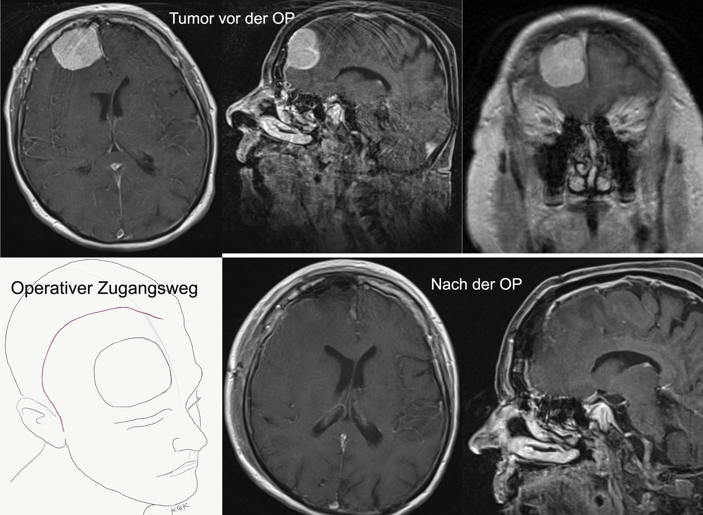
Example 2: The patient is a 74-year-old right-handed gentleman—a devoted father and grandfather in a large, affluent family. Despite repeated substantial financial losses, he had developed an uncharacteristic compulsion for gambling over recent months. His family noticed progressive irritability, uncharacteristic aggression, and inappropriate, socially uninhibited language during conversations. One day, the patient suffered a major generalized epileptic seizure with transient loss of consciousness and was brought to the emergency department. Cranial MRI revealed a large, smoothly circumscribed, homogenously contrast-enhancing tumor in the right non-dominant frontal lobe with surrounding vasogenic edema. In view of the tumor's size and mass effect, the patient underwent microneurosurgical resection. The tumor was completely removed. Following surgery, the patient's neuropsychological symptoms resolved and he was able to return to his normal family life. Neuropathological evaluation confirmed a WHO Grade I meningioma (a benign diagnosis). Because complete resection was achieved and the tumor was benign, no adjuvant therapy was required. The patient is monitored through regular clinical and MRI follow-ups. Nota bene: The clinical course in your specific case may differ from the example described here.
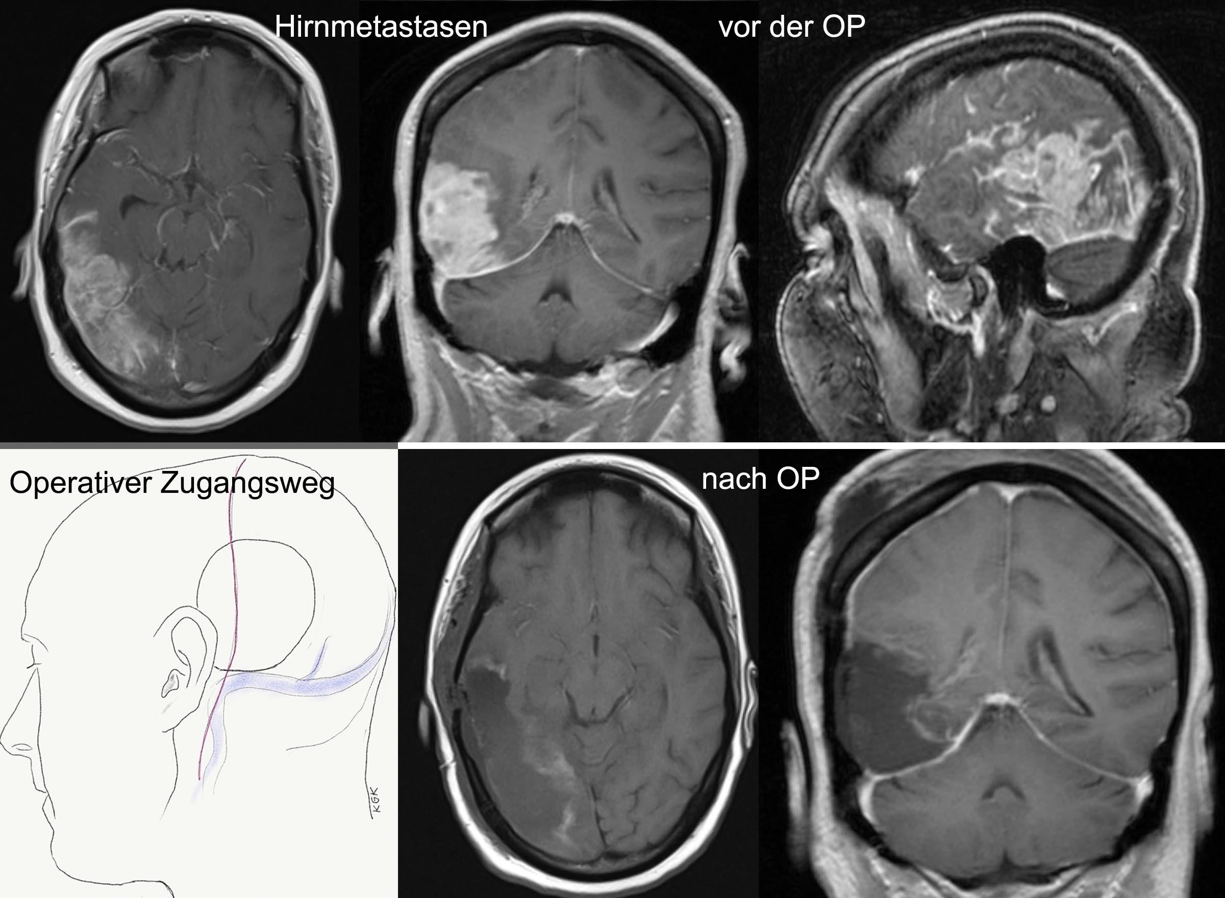
Example 3: A 36-year-old right-handed woman suffered sudden loss of consciousness. One year prior, she had undergone surgical excision of a breast malignancy followed by adjuvant chemotherapy. On this occasion, she lost consciousness for approximately 30 minutes, regaining consciousness spontaneously without intervention—in all likelihood representing an epileptic seizure with postictal state. Diagnostic imaging revealed a large mass in the right cerebral hemisphere situated between the sensory cortex and the visual cortex, alongside a second smaller tumor in the cerebellum. Through an osteoplastic craniotomy, the dominant cerebral tumor was resected under the operating microscope utilizing intraoperative neuronavigation, intraoperative real-time ultrasonography, and an ultrasonic aspirator. Neuropathological examination confirmed the expected diagnosis of metastatic breast adenocarcinoma. Postoperative adjuvant chemotherapy and whole-brain radiation therapy (WBRT) were administered, supplemented by patient-specific targeted immunotherapy with monoclonal antibodies. Nota bene: The clinical course in your specific case may differ from the example described here.