
Tumors of the Pituitary Gland:
Pituitary tumors (pituitary adenomas) occur in approximately 2–3 per 100,000 individuals per year. The vast majority of pituitary tumors are hormone-producing adenomas.
Hormonally active, prolactin-secreting tumors (prolactinomas) frequently manifest through menstrual cycle irregularities (in women) and loss of libido or hypogonadism (in men).
Growth hormone (GH)-secreting tumors cause acromegaly (characterized by enlargement of the hands, feet, jaw, and tongue, deepening of the voice, and coarse facial features).
Corticotropin (ACTH)-secreting pituitary tumors cause Cushing's disease ("moon face", doughy soft tissues, central/truncal adiposity with slender limbs, and arterial hypertension).
Hormonally inactive (non-functioning) pituitary tumors typically manifest through mass effect and compression of adjacent anatomical structures, particularly the optic chiasm (causing visual disturbances, visual field defects such as bitemporal hemianopsia, and progressive vision loss).
The choice of treatment depends on the size, endocrine profile, and anatomical extension of the pituitary tumor. The optimal therapy is complete surgical excision of the tumor tissue. In the vast majority of cases, endocrine metabolic balance normalizes fully following surgery, and symptoms resolve. The modern standard of care and preferred surgical route is the fully endoscopic transnasal-transsphenoidal approach through one or both nostrils. In the vast majority of patients, the tumor can be resected completely and gently via this minimally invasive transnasal corridor.
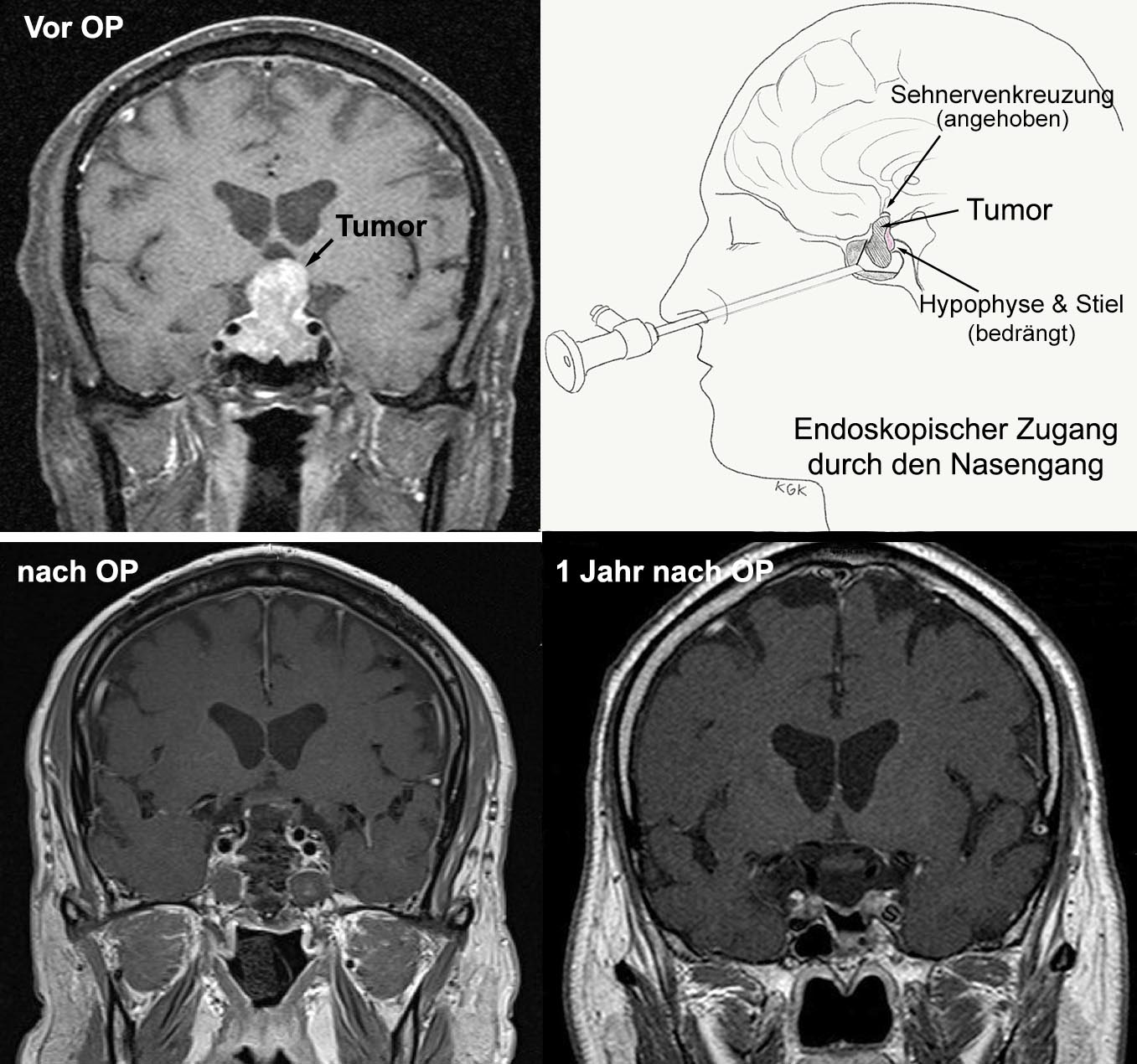
Example 4: A 50-year-old patient noticed that over the course of approximately 18 months, his shoe and glove sizes were increasing rapidly. His voice had deepened and his facial appearance had noticeably changed—typical features of acromegaly. Over the preceding month, his peripheral vision on the left side deteriorated, causing him to repeatedly bump into door frames and objects on his left. Endocrinological diagnostic workup revealed marked elevations in IGF-1 and prolactin levels. Ophthalmological evaluation documented significant visual field constriction on the left side (temporal hemianopsia). High-resolution sellar MRI confirmed a pituitary macroadenoma with suprasellar elevation of the optic chiasm. Surgical intervention was recommended. Following comprehensive endocrine and anesthetic preparation, a fully endoscopic transnasal-transsphenoidal tumor resection was performed. The macroadenoma was excised completely along with its capsule. Postoperatively, the patient received temporary hydrocortisone replacement therapy, which was successfully tapered and discontinued within three months. Visual fields improved promptly and normalized within one week after surgery. Shoe and glove sizes stabilized without further progression. Neuropathological analysis confirmed a hormonally active pituitary macroadenoma. Because complete tumor clearance was achieved, no additional therapy was required. Long-term follow-up over several years revealed no tumor recurrence. Nota bene: The clinical course in your specific case may differ from the example described here.