
Tumors of the Spine and Spinal Cord:
Spinal tumors may:
- affect solely the vertebral bones (extradural/osseous: metastases, lymphoma, plasmacytoma / multiple myeloma)
- be located within the spinal canal without infiltrating the spinal dura mater (extradural: epidural metastases, cyst, exophytic meningioma)
- be situated within the spinal dura mater without infiltrating the spinal cord parenchyma itself (intradural-extramedullary: meningioma, schwannoma)
- arise directly from the neural tissue of the spinal cord (intramedullary: ependymoma, astrocytoma, glioma)
Depending on tumor entity, histological grade, anatomical extent, and invasiveness, spinal tumors are treated surgically, chemotherapeutically, and/or radiotherapeutically, either individually or in multimodal combination. During resection and adjuvant treatment of spinal tumors, preserving and restoring spinal biomechanical stability is just as critical as oncological tumor clearance.
Modern spinal neurosurgery utilizes sophisticated specialized modalities: multimodal intraoperative neurophysiological monitoring (IONM) to continuously safeguard neural pathways, spinal neuronavigation, microscopic, endoscopic, and exoscopic visualization techniques, and instrumented spinal stabilization with pedicle screw-rod systems.
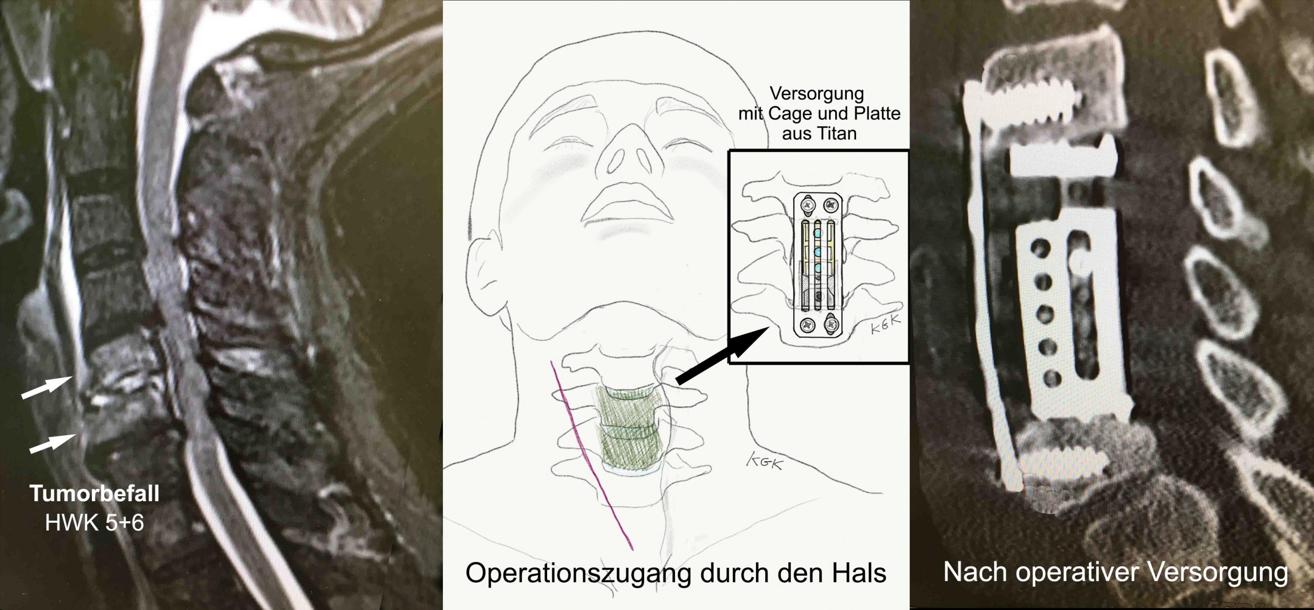
Example 8: A 58-year-old male patient presented with neck pain radiating into both arms that had persisted for several months. Analgesic medications and physiotherapy provided no relief. Outpatient spinal injection therapies had also proven ineffective. [Medical history: The patient had been treated 10 years earlier for prostate adenocarcinoma with curative outcome]. Because of the persistent, progressive pain, an MRI of the cervical spine was performed. Imaging demonstrated extensive tumor destruction of cervical vertebrae C5 and C6. Following thorough interdisciplinary preparation, surgery was performed: the affected, collapsed vertebral bodies were resected via an anterior cervical approach (corpectomy). The resulting vertebral defect was reconstructed with an expandable titanium cage, and spinal stability was secured with an anterior plate osteosynthesis spanning C4 to C7. Following interdisciplinary tumor board review, the patient underwent targeted radiation therapy and chemotherapy. Excellent local tumor control was successfully established. Nota bene: The course of disease in your specific case may differ from the clinical example described here.
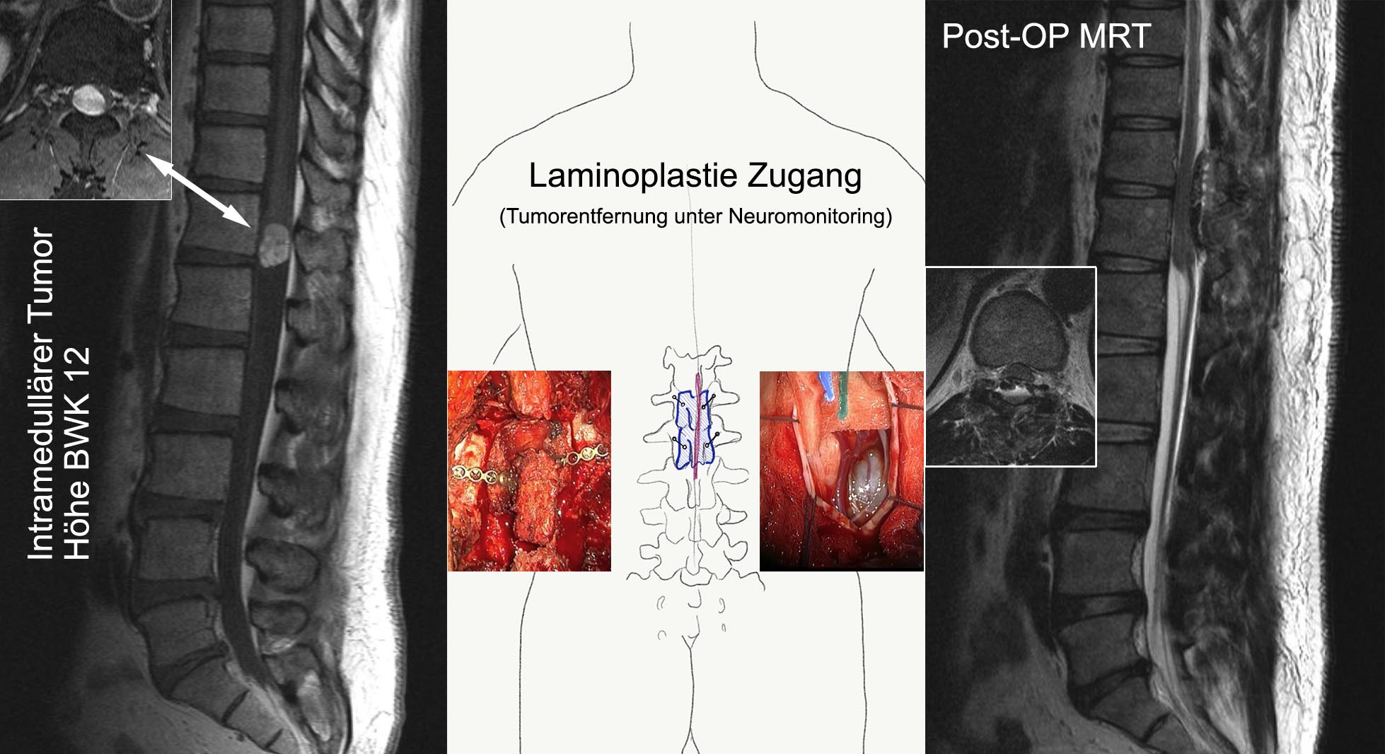
Example 9: A 34-year-old software engineer experienced involuntary shaking and clonus in both legs for approximately one year. Episodes lasted roughly 3 minutes and occurred more frequently at night. He initially attributed these symptoms to long hours of sitting at work. Over the preceding 4 months, severe back pain emerged, accompanied by bladder emptying difficulties (urinary dysfunction). MRI studies revealed an intradural tumor at the thoracolumbar junction (T12–L1). The lesion was sharply demarcated and encapsulated, strongly suggesting a benign pathology. Following preoperative planning, an osteoplastic laminoplasty approach was performed: the posterior bony laminar arches were removed en bloc and subsequently refixed with microplates and screws after tumor resection, thus preventing any post-laminectomy kyphotic deformity or loss of spinal stability. The tumor resection was carried out microsurgically under continuous intraoperative neuromonitoring to safeguard neural integrity. The lesion was completely resected (gross-total resection). Neuropathological analysis confirmed a Schwannoma (WHO Grade I) — a benign nerve sheath tumor, completely excised. Consequently, no adjuvant therapy was needed. Postoperatively, the patient showed no neurological deficits. Follow-up MRI scans up to four years post-surgery documented zero tumor recurrence. Nota bene: The course of disease in your specific case may differ from the clinical example described here.