Trigeminal Neuralgia, Hemifacial Spasm, and Neurovascular Compression Syndromes
Trigeminal Neuralgia
The trigeminal nerve (cranial nerve V) is responsible for facial sensation and divides into three major sensory branches: (1) the ophthalmic nerve (V1: forehead, eye, and upper eyelid), (2) the maxillary nerve (V2: cheek, upper jaw, upper teeth, and upper lip), and (3) the mandibular nerve (V3: lower jaw, lower teeth, lower lip, and chin). In addition, the trigeminal nerve supplies motor innervation to the muscles of mastication (including the masseter and temporalis muscles).
In trigeminal neuralgia, typically one or two of these sensory branches are affected. The condition is characterized by paroxysmal, lightning-like, excruciating electrical shock-like pain attacks, often provoked by minimal trigger stimuli (such as chewing, speaking, washing the face, brushing teeth, light touch, or a cool breeze). Trigeminal neuralgia inflicts an immense degree of suffering, profoundly undermining daily functioning and emotional well-being.
Hemifacial Spasm
The facial nerve (cranial nerve VII) provides voluntary motor control to the mimetic musculature of the face and divides into five main branches: (1) temporal/frontal and zygomatic branches (forehead and eye closure), (2) buccal branches (midface and upper lip elevation), (3) marginal mandibular branch (mouth corner movements and lower lip), (4) mandibular branches, and (5) cervical branch (platysma). The facial nerve also conveys autonomic parasympathetic fibers controlling lacrimal (tear) and salivary gland secretion.
Like trigeminal neuralgia, hemifacial spasm is a profoundly disabling condition. Chronic mechanical irritation and hyperexcitability of the facial nerve generate involuntary, uncontrollable twitching and clonic spasms of the ipsilateral facial musculature. In certain cases, muscle spasms can be accompanied by intense episodic pain (tic douloureux; painful facial spasms).
Causes of Trigeminal Neuralgia, Hemifacial Spasm, and Related Cranial Nerve Conditions:
In the overwhelming majority of patients (approaching 100%), a neurovascular conflict at the root entry or exit zone (REZ) of the cranial nerve in the cerebellopontine angle / posterior cranial fossa is the underlying etiology. Chronic mechanical compression by an elongated or looping blood vessel (typically an artery, occasionally a vein) against the brainstem root of the nerve—combined with relentless arterial pulsation over decades—leads to localized demyelination (focal breakdown of the nerve's protective myelin sheath). This microstructural injury creates pathological ephaptic transmission ("cross-talk" and electrical short-circuiting between adjacent nerve fibers), resulting in paroxysmal symptoms.
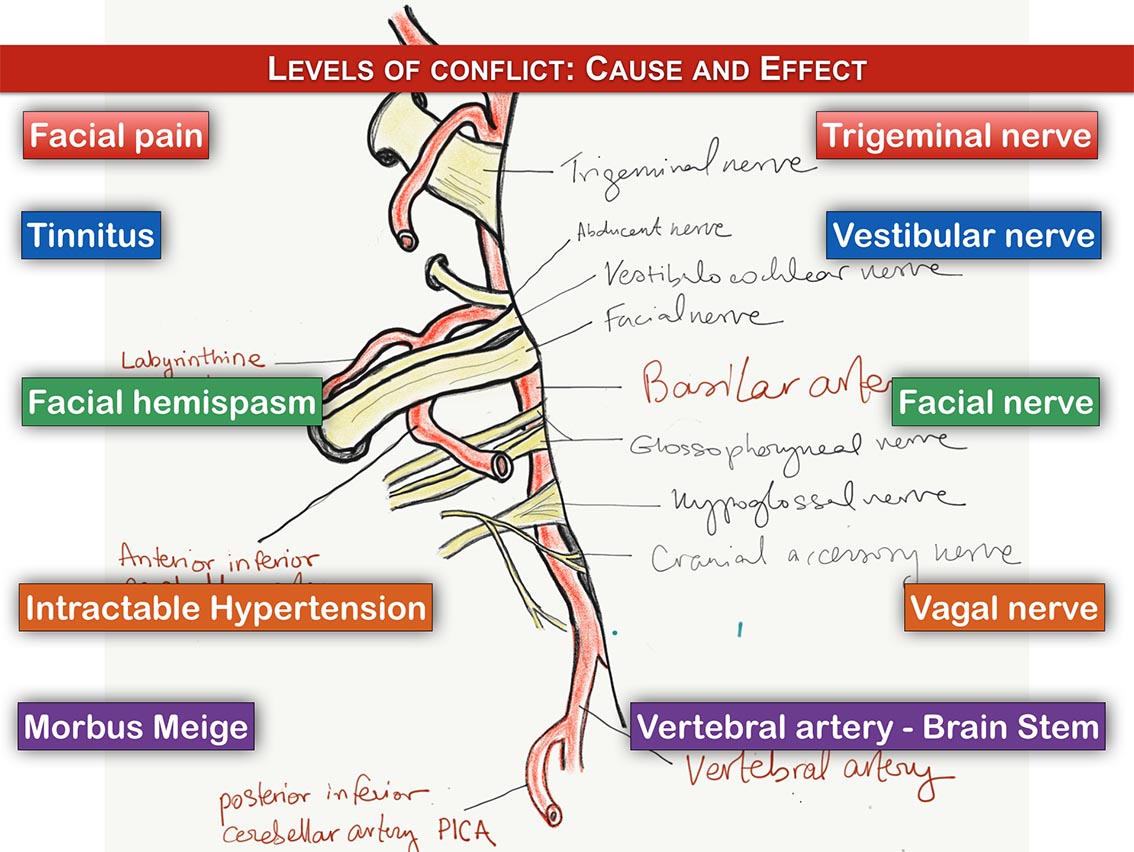
- When the facial nerve (CN VII) is affected: Hemifacial spasm develops
- When the trigeminal nerve (CN V) is affected: Trigeminal neuralgia develops
- When the vagus nerve (CN X) is affected: Treatment-refractory neurogenic hypertension can arise
- When the vestibulocochlear nerve (CN VIII) is affected: Intractable tinnitus or disabling vertigo may occur
- When the glossopharyngeal nerve (CN IX) is affected: Glossopharyngeal neuralgia or intractable involuntary hiccups develop
Principles of Treatment:
The first-line therapy for neurovascular cranial nerve compression syndromes is conservative and pharmacological. Centrally acting anticonvulsant and membrane-stabilizing medications (such as carbamazepine, oxcarbazepine, or gabapentin) are titrated to suppress involuntary, ectopic nerve impulses and electrical discharges. In a significant proportion of patients, medical management provides initial symptom relief.
Pharmacological treatment should ideally be supported by psychological counseling, pain management behavioral therapy, and autogenic relaxation training. These modalities help patients systematically recognize and avoid specific trigger factors, while building effective coping mechanisms to overcome pain-related fear, anticipatory anxiety, and pain memory.
Over time, however, many patients experience waning drug efficacy, breakthrough pain crises, or debilitating pharmaceutical adverse effects (such as marked lethargy, cognitive dulling, dizziness, or hepatic toxicity), necessitating medical discontinuation.
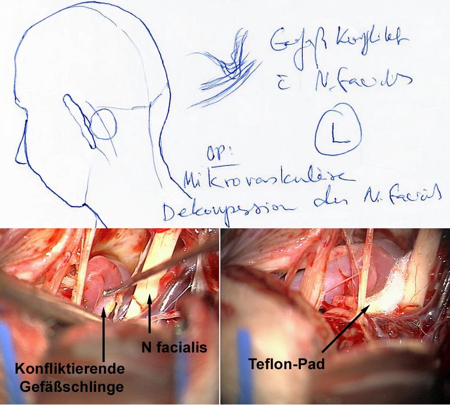
In such refractory or intolerant cases, neurosurgical intervention is indicated. The causal treatment of choice is microvascular decompression (MVD, Janetta procedure). Through a keyhole retrosigmoid craniotomy, the compressing vascular loop is gently mobilized away from the nerve root exit zone under the operating microscope, and an inert Teflon sponge or felt spacer is interposed to permanently eliminate mechanical pulsatile contact. Because centralized pain memory and neural hyperpathia may transiently persist even after mechanical relief, surgical treatment is integrated with thoughtful medical tapering and supportive care.
High-precision stereotactic radiosurgery (such as CyberKnife® or Gamma Knife®) provides a proven, non-invasive alternative for selected patients who are unsuitable for or wish to avoid microsurgery. Targeted, millimeter-precise radiation induces controlled, focal fibrosis (scarring) within the nerve root, dampening aberrant electrical hyper-conduction. The primary advantage of radiosurgery is that it is performed completely non-invasively, without requiring posterior fossa craniotomy.
Nota bene: The information provided above may not directly reflect your individual clinical circumstances. Every patient must be evaluated individually to formulate a tailored, optimal diagnostic and therapeutic strategy.