
Hemifacial Spasm (Spasmus Hemifacialis)
The facial nerve (cranial nerve VII) provides voluntary motor innervation to the mimetic musculature of each side of the face and divides into five main terminal branches: (1) temporal/frontal and zygomatic branches (forehead and periorbital muscles), (2) buccal branches (midface and upper lip muscles), (3) marginal mandibular branch (oral commissure and lower lip excursion), (4) mandibular branches, and (5) cervical branch (platysma). In addition, the facial nerve carries autonomic parasympathetic fibers controlling lacrimation (tearing) and salivation.
Much like trigeminal neuralgia, hemifacial spasm is a deeply debilitating condition. Persistent mechanical irritation and hyper-excitability of the facial nerve produce involuntary, uncontrollable twitching and clonic contractions of the facial muscles, typically beginning around the orbicularis oculi (eyelid twitching) and gradually spreading across the entire half of the face. In some cases, the spasmodic contractions are accompanied by severe episodic pain (tic douloureux; painful facial spasms).
Causes of Hemifacial Spasm, Trigeminal Neuralgia, and Related Conditions:
In the vast majority of patients (approaching 100%), high-resolution MRI reveals a neurovascular conflict at the root exit zone (REZ) of the cranial nerve in the cerebellopontine angle / posterior fossa. The mechanical compression of an arterial loop (such as the anterior inferior cerebellar artery [AICA], posterior inferior cerebellar artery [PICA], or vertebral artery) against the brainstem root of the facial nerve—along with constant pulsatile trauma over decades—leads to localized demyelination (thinning of the protective myelin sheath). This microstructural breakdown leads to ephaptic transmission (electrical "short circuits" among neighboring nerve fibers), driving the involuntary paroxysmal spasms.
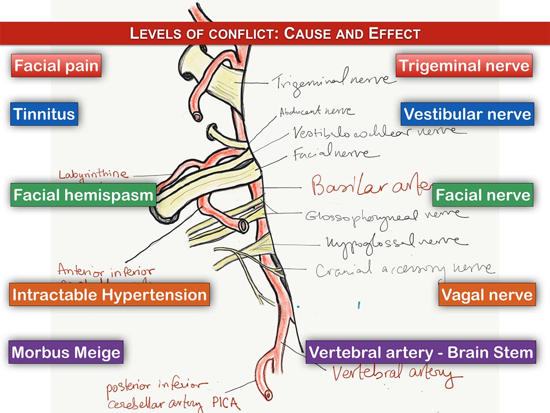
- When the facial nerve (CN VII) is compressed: Hemifacial spasm develops
- When the trigeminal nerve (CN V) is compressed: Trigeminal neuralgia develops
- When the glossopharyngeal nerve (CN IX) is compressed: Glossopharyngeal neuralgia or intractable hiccups develop
- When the vagus nerve (CN X) is involved: Refractory neurogenic hypertension can arise
- When the vestibulocochlear nerve (CN VIII) is compressed: Intractable tinnitus or disabling vertigo may occur
Principles of Treatment:
The first-line conservative treatment for cranial nerve compression syndromes often involves medications, such as membrane-stabilizing and anticonvulsant drugs (e.g., carbamazepine, gabapentin, or baclofen), which suppress involuntary, ectopic electrical discharges. In mild or early hemifacial spasm, regular local injections of botulinum toxin (Botox) can also temporarily alleviate spasms by blocking neuromuscular transmission at the periphery.
Medical therapies can be supplemented by stress-reduction modalities, psychological counseling, and autogenic relaxation training, which assist patients in identifying stress-related spasm triggers and developing effective coping strategies.
However, medical therapies do not resolve the anatomical cause, and patients frequently develop tolerance, diminishing benefit, or intolerable systemic side effects over time.
In these refractory or progressive cases, causal neurosurgical treatment is indicated. The definitive standard of care is microvascular decompression (MVD, Janetta procedure). Performed via a minimally invasive retrosigmoid keyhole craniotomy under continuous neurophysiological intraoperative monitoring (IOM), the compressing vascular loop is gently mobilized away from the facial nerve root exit zone, and an inert Teflon felt spacer is placed between the vessel and the brainstem to permanently eliminate mechanical pulsatile contact.
High-precision stereotactic radiosurgery (such as CyberKnife® or Gamma Knife®) serves as an alternative non-invasive option for selected patients who are poor surgical candidates. Focused radiation induces localized micro-fibrosis (scarring) at the nerve root, mitigating pathological electrical hyper-conduction without requiring open surgery or craniotomy.
Nota bene: The information provided above may not directly reflect your individual clinical circumstances. Every patient must be evaluated individually to establish the optimal, safest therapeutic approach.