
Vertebral Fractures:
Spinal fractures typically result from traumatic injuries; however, pathological fractures also frequently occur in the context of osteoporosis and neoplastic disease (bone metastases or primary tumors).
Unstable vertebral fractures pose a severe threat to neurological function (such as paraplegia or quadriplegia) and require prompt surgical management aimed at spinal stabilization and decompression. The treatment options for osteoporotic and tumor-related fractures depend upon the extent and biomechanics of the lesion. For example, if there is solely an osteoporotic reduction in vertebral body height without compromised spinal canal or neural compression, percutaneous injection of bone cement into the vertebral body (balloon kyphoplasty or vertebroplasty) is completely sufficient. Depending on the tumor entity, emergency radiation therapy may be pursued to establish local tissue consolidation. When neurological function is compromised or threatened, surgical stabilization combined with simultaneous decompression of the spinal cord is imperative.
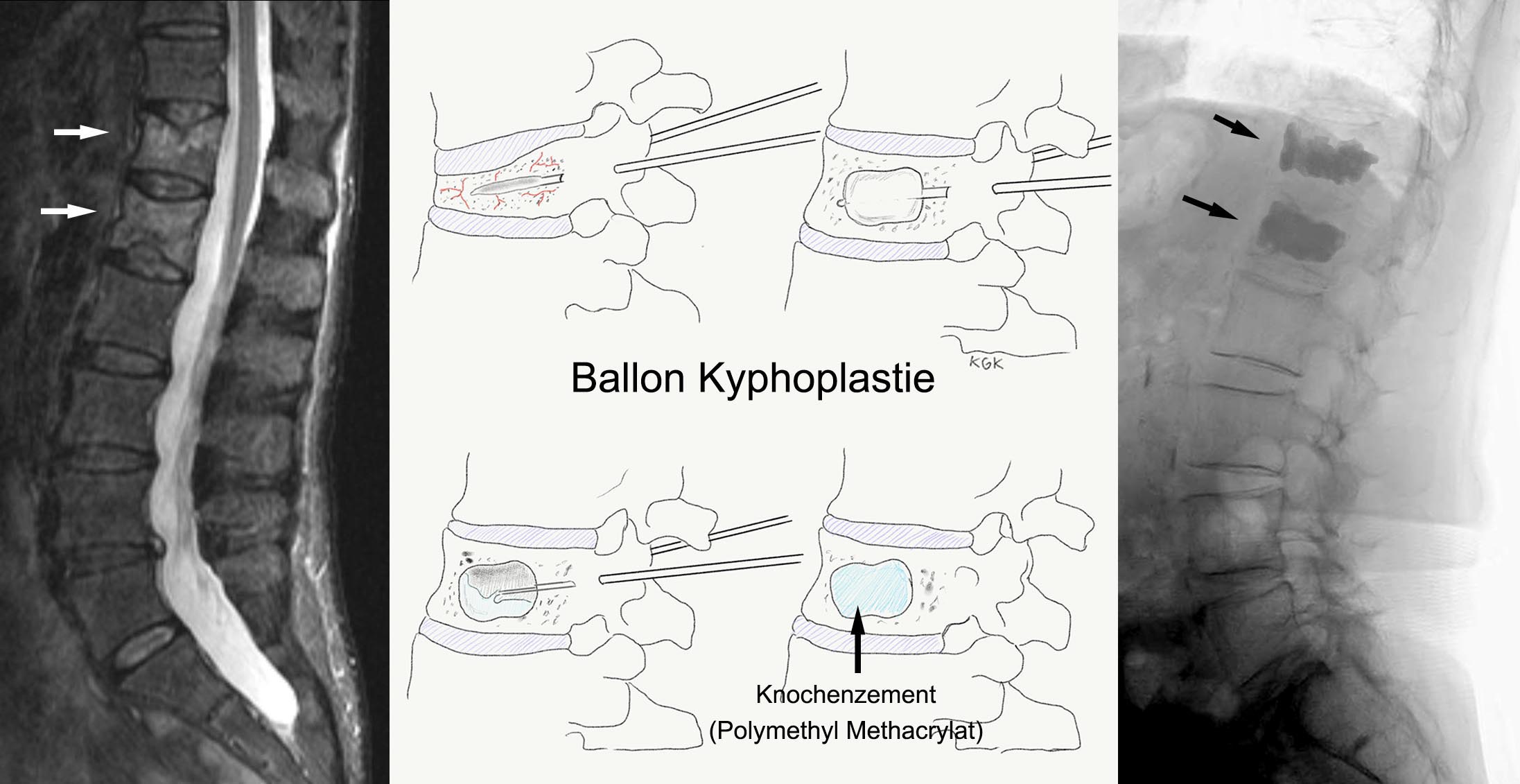
Example 1: An 82-year-old female patient slipped in the bathroom, sustaining a severe back contusion. The severe back pain failed to improve even after a week of conservative medication, prompting her to see a specialist. An X-ray demonstrated marked height loss of the 12th thoracic vertebra (T12) and 1st lumbar vertebra (L1). Relevant medical history included osteoporosis diagnosed 10 years earlier, for which she had been receiving ongoing treatment. An MRI of the spine revealed acute pathological compression fractures of both T12 and L1. In view of her intractable pain, a balloon kyphoplasty was performed under general anesthesia. Following the procedure, the pain decreased significantly and the patient was discharged home the next day without mobility restrictions. Nota bene: The course of disease in your specific case may differ from the clinical example described here.
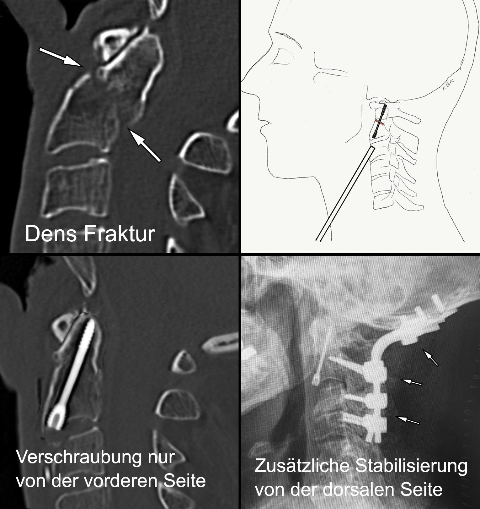
Example 2: Following an episode of vertigo, an 84-year-old male patient fell backward, striking the occiput against a radiator and sustaining a severe cervical hyperextension/whiplash injury. A CT scan revealed an odontoid fracture (Dens axis fracture, Type I – IIa). In such an uncomplicated fracture configuration, surgical management consists of direct anterior osteosynthesis using a single cannulated lag screw placed from the anterior cervical approach. This screw anatomically realigns and securely fixes the dens process. In more complex fractures (Types IIb and III), additional (or stand-alone) posterior instrumentation and fusion is indicated. With such fusion procedures, cervical rotation is reduced to a certain degree. Nota bene: The course of disease in your specific case may differ from the clinical example described here.
In some traumatic injuries, the bony spinal structures remain intact, yet severe ligamentous disruptions occur. Ligamentous tears can cause profound spinal instability that is potentially life-threatening and functionally catastrophic. In these situations, prompt external spinal immobilization for patient transport followed by swift surgical stabilization and reconstruction is indicated.
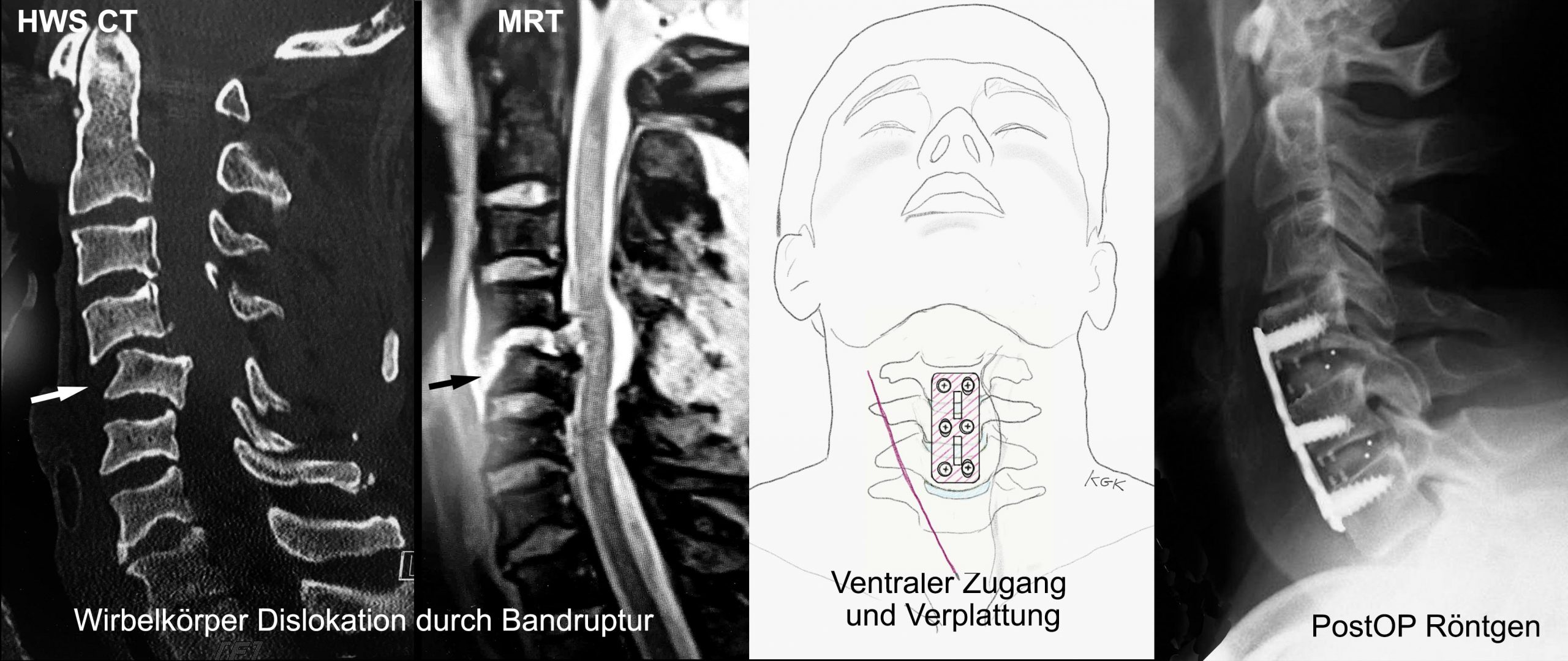
Example 3: A 38-year-old equestrian sustained a high-energy fall from horseback, striking his head and experiencing the immediate sensation of a severe cervical fracture. He initially felt an intense electric shock sensation radiating into his left arm and leg, followed by profound warmth. Upon arrival of emergency medical services, he was unable to move his left leg, and could only move his right leg with difficulty. Once the emergency physician applied a rigid cervical collar for immobilization, sensory perception in his leg began to recover. An emergency CT scan revealed a complete posterior dislocation of the 5th cervical vertebra (C5) caused by traumatic rupture of the spinal ligaments. Following swift preoperative preparation, surgery was carried out: anatomic reduction and anterior plate osteosynthesis from C4 to C6 were performed. Despite stabilization spanning three vertebrae, cervical spine mobility remained widely preserved thanks to the adjacent mobile segments. Given the modest preoperative deficits, his young age, and immediate operative management, the patient was able to return to his personal and professional life without restrictions following rehabilitation. Nota bene: The course of disease in your specific case may differ from the clinical example described here.